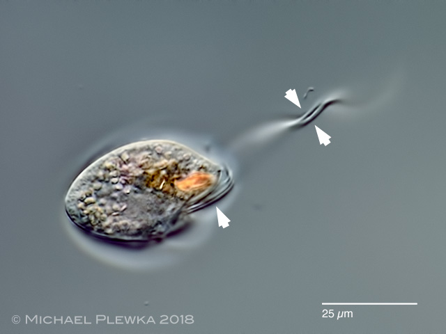
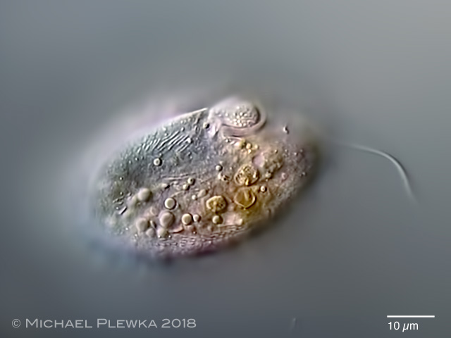
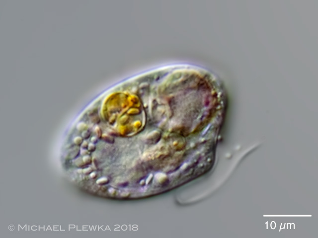
| 1.Oxyrrhis marina: a heterotroph (phagotroph) Dinoflagellate. The arrows point to the two flagella which insert ventrally and sometimes are orientated parallel to each other. |
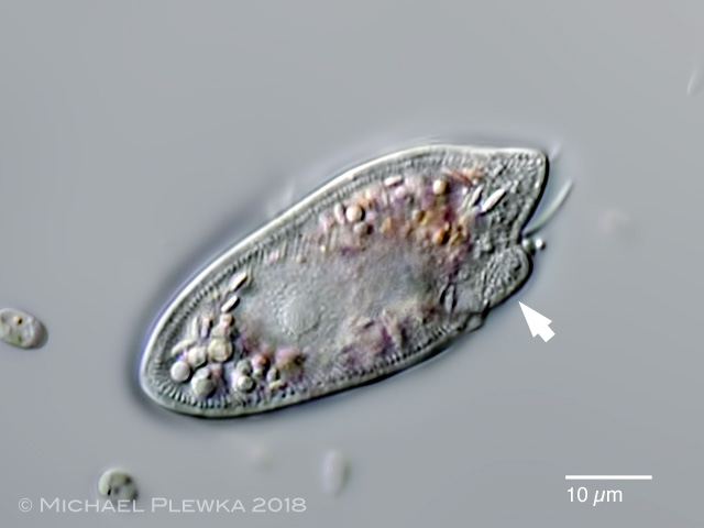 |
| 2.Oxyrrhis marina: focus plane on the nucleus. The cortex shows a layer of ?trichocysts? or ?scales?. The arrow points to the ventral bulge. O. marina has no contractile vacuole. |
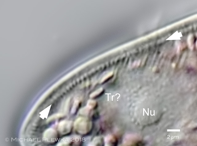 |
| 3.Oxyrrhis marina: crop of the above image. The arrowheads mark a dense layer of fusiform structures below the outer laye of the cortexr. The oval structures might be trichocysts (Tr). Nu: nucleolus. |
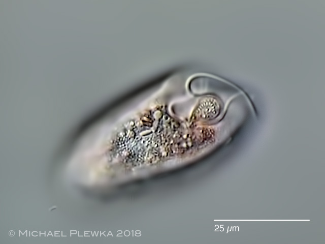 |
| 4.Oxyrrhis marina: focus plane on the tentacle/ ventral bulge |
 |
| 5.Oxyrrhis marina: another specimen with focus plane on the tentacle/ ventral bulge. |
 |
 |
| 6/7.Oxyrrhis marina: two images from a video showing a specimen which had engulfed a cyanobacterium, presumably a Synechocystis sp., and was trapped by the coverslide. |
 |
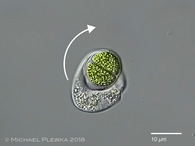
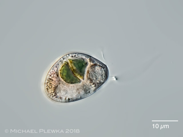
| 8.Oxyrrhis marina: The arrow points to the stigma of an engulfed chlorophyte. |
 |
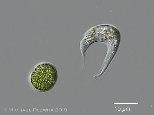
| 9.Oxyrrhis marina: with another prey. The cells of Oxyrrhis marina appear pinkish due to Rhodopsin. |
 |
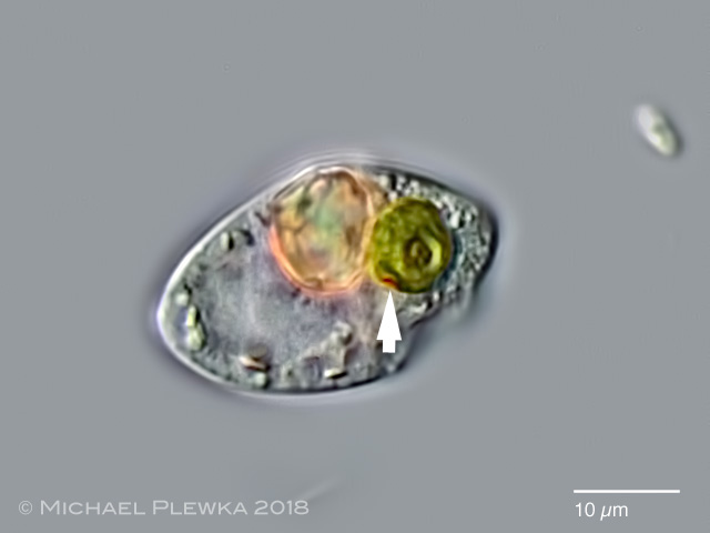
| 10.Oxyrrhis marina: with a partly digested cyanobacterium. |
| |
| Location: Porspoder, Brittany, France, tide pool |
| Habitat: plankton |
| Date: 08.08.2018 |
| |
|
| |
|